To save an image onto your computer, right click it and select "save as".
All images are copyright Magscope. They are released under the
Creative Commons Licence CC BY-NC-ND 3.0
You may download and use these images for non-profit purposes, but the notices on each image must remain intact.
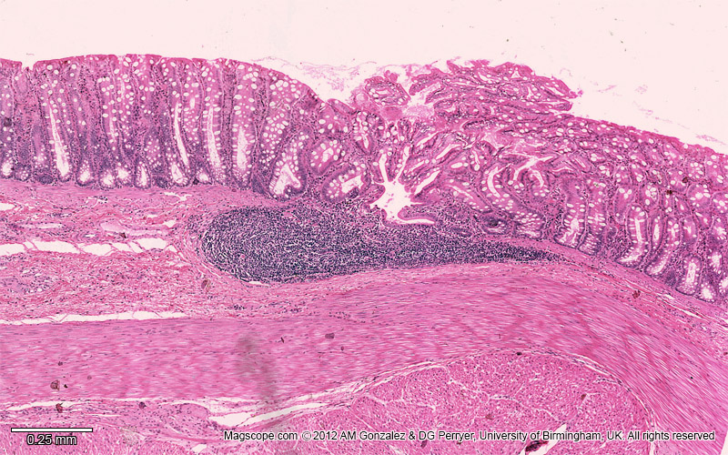
Micrograph of the rectum illustrating the epithelium, lamina propria, submucosa and muscularis externa
Low magnification image of the rectum illustrating the simple columnar epithelium with numeorus goblet cells in the mucosa, a solitary lymph node in the lamina propria and submucosa and the muscularis externa
Disability awareness and educational equity: This image has been optimised for red-green colour blind observers who are often unable to differentiate the colours in histological slides, using methods described by Professors Landini and Perryer here.
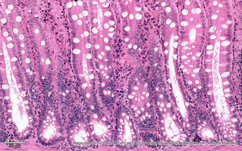
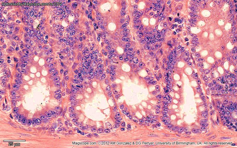
Micrograph of the simple columnar epithelium of the rectum containing numerous goblets cells
Disability awareness and educational equity: This image has been optimised for red-green colour blind observers who are often unable to differentiate the colours in histological slides, using methods described by Professors Landini and Perryer here.
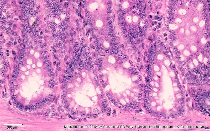
High magnification image of the basal portion of the rectal epithelium illustrating the tall single columnar epithelial cells and goblet cells
Disability awareness and educational equity: This image has been optimised for red-green colour blind observers who are often unable to differentiate the colours in histological slides, using methods described by Professors Landini and Perryer here.
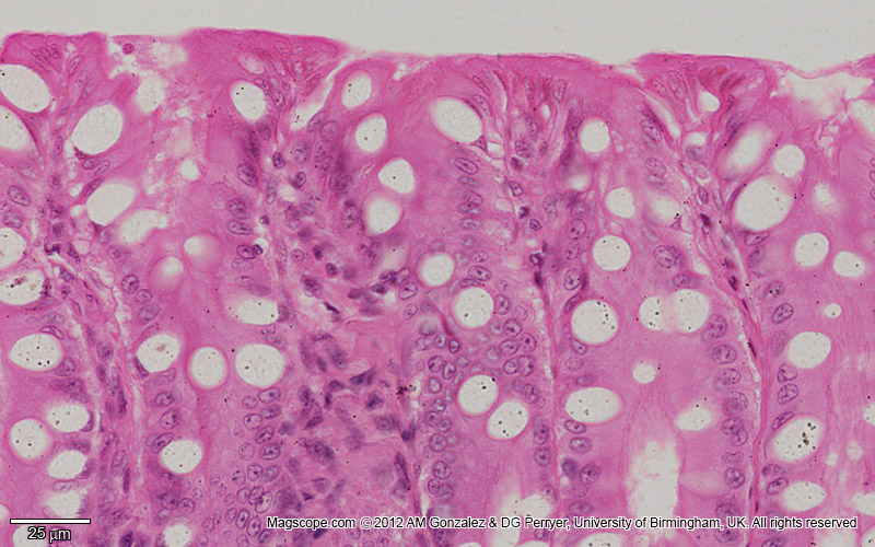
High magnification image of the surface portion of the rectal epithelium illustrating the tall single columnar epithelial cells and goblet cells.
Disability awareness and educational equity: This image has been optimised for red-green colour blind observers who are often unable to differentiate the colours in histological slides, using methods described by Professors Landini and Perryer here.
Funding for the Slide Bank was generously provided by

 All images are copyright Magscope. They are released under the
Creative Commons Licence CC BY-NC-ND 3.0
All images are copyright Magscope. They are released under the
Creative Commons Licence CC BY-NC-ND 3.0