
Skin - Pigmented
Micrograph of a skin section illustrating the thick keratinised pigmented epithelium of the epidermis and subjacent dermis
Magscope Virtual Microscope
Teaching Images
4 images
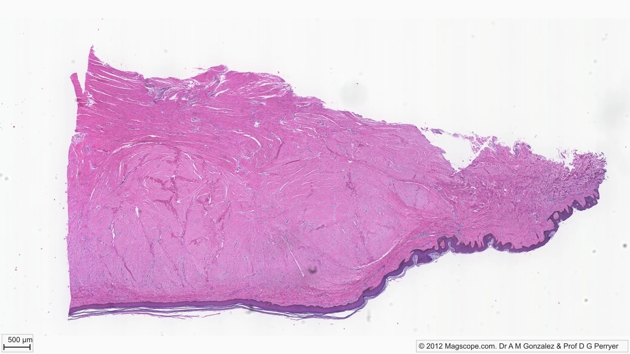
Pigmented skin overview. Dense collagenous dermis fills most of the section, with a dark keratinised epidermal band and folded rete ridges along the surface.
Melanin and keratin. Brown melanin is concentrated near basal and lower spinous keratinocytes, while partly lifted stratum corneum forms pale keratin sheets at the surface.
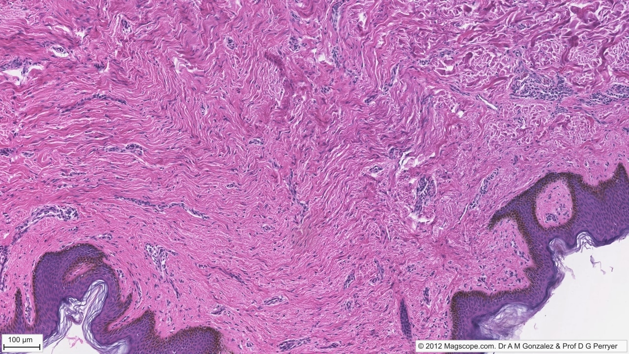
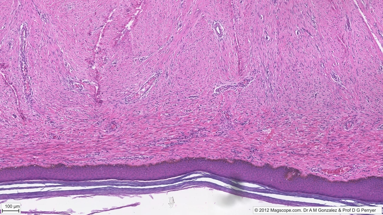
Folded pigmented epidermis. Deep rete ridges alternate with dermal papillae, while dense irregular connective tissue contains wavy collagen bundles, vessels and fibroblast nuclei.
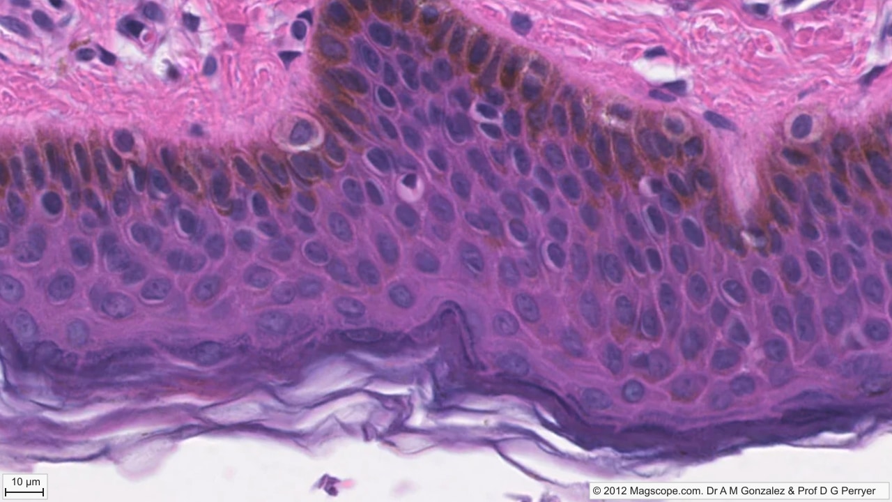
Pigment close-up. Brown melanin granules are prominent in basal and lower suprabasal keratinocytes near the dermal junction, while lifted stratum corneum marks the free surface.
Images © Magscope. Released under CC BY-NC-ND 3.0 for non-commercial use only. Images must remain unmodified and all copyright/licence notices must remain intact. Commercial use, notice removal, resale or redistribution requires a separate Magscope licence.